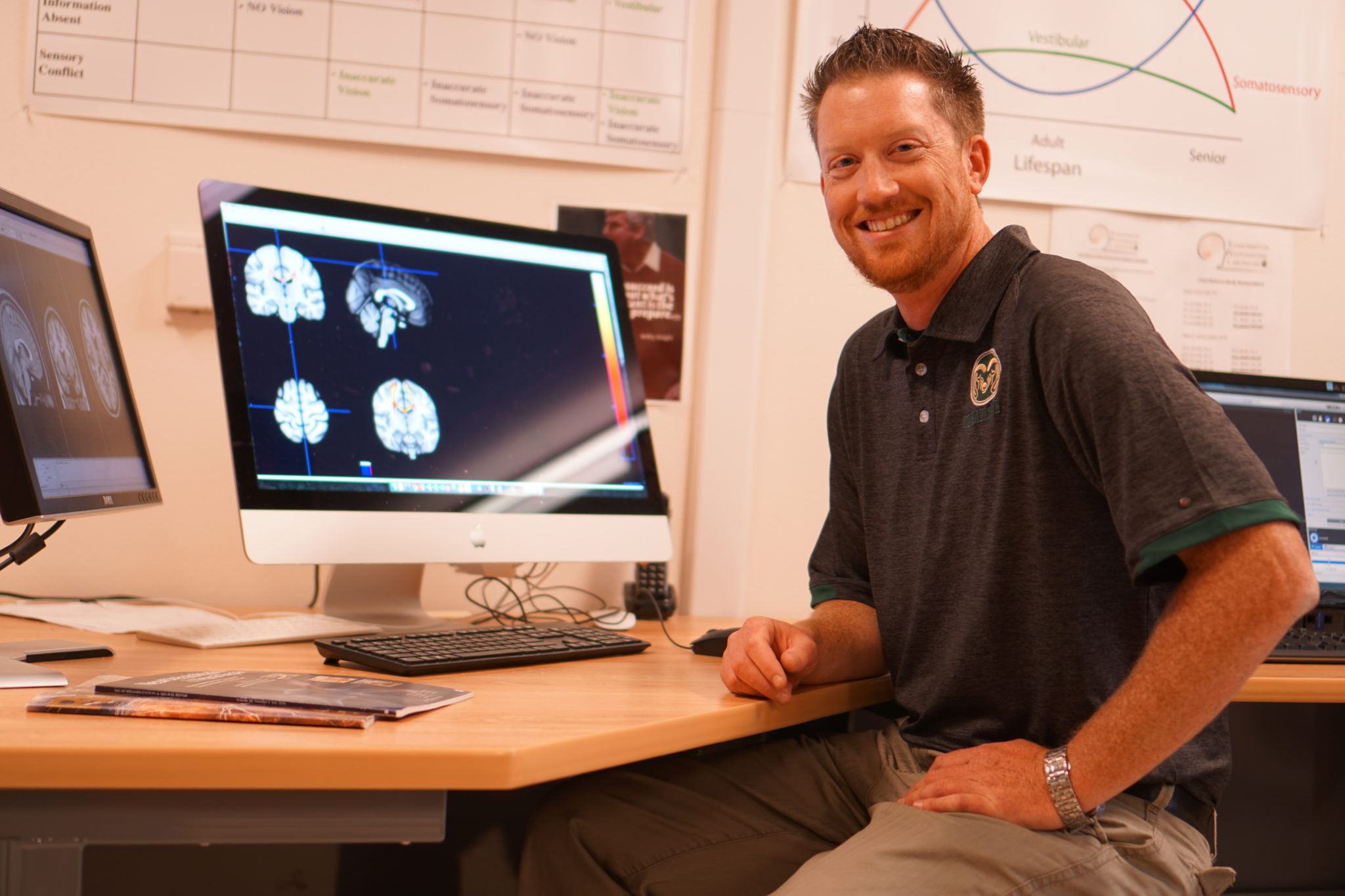
Walking with balance and coordination is something most of us do every day without thinking much about it. For individuals with multiple sclerosis, it’s a different story. A Colorado State University researcher has received funding to investigate how the brain connects to the body in individuals with MS.
Brett Fling, assistant professor in the Department of Health and Exercise Science, is the primary investigator in a study titled “Two legs, one brain…” which has received a $200,000 grant over the next three years from the Dana Foundation’s neuroimaging program. Fling heads up CSU’s Sensorimotor Neuroimaging Laboratory. His research team hopes to find a way to target gait rehabilitation.
MS and the brain
In individuals with multiple sclerosis, the brain’s white matter is impaired. Think of white matter as the electrical wiring that carries messages from point A to point B. For people with MS, the myelin sheath that keeps the wires insulated is lost and the messages have trouble getting from A to B. This causes the messages to lose speed and accuracy, impairing motor skills.
Each individual with MS has distinct issues, and not everyone has the same symptoms. For this project, Fling and his team will be studying people with MS who are still able to walk on their own. People with MS tend to have a good side and a bad side, where the individuals have difficulty controlling one side of their body. Scientists don’t understand why these individuals have a more-affected and less-affected side.
Complex interactions
“We have a pretty good idea of how our brain controls the complex interactions that take place when we coordinate our hands,” explained Fling. “Think about buttoning a shirt or tying your shoes; each hand is doing something different and independent in time and space, but they are coordinated together perfectly.”
“It’s a lot more complicated to see how our brain controls walking and balance,” Fling continued. “We are constantly having to maintain balance so we don’t fall over. It’s usually an automated process that we don’t think about. Once an individual develops impairments such as MS, the automatic correction is lost. Walking becomes a much more mentally taxing activity to maintain gait and balance. We really don’t know how our brain does it.”

Research study
Fling and his team will be using different approaches to look at brain structure to see how the brain is failing to communicate correctly. They hope to use this information to understand why individuals with multiple sclerosis have gait and balance disorders.
In their study, Fling’s team will focus on a large bundle of white matter, called the corpus callosum, which connects the two sides of the brain. The corpus callosum is used in hand dexterity and coordination, and the research team will be focusing on this brain structure while studying balance coordination.
This research will use MRI as well as Transcranial Magnetic Stimulation to look at how the two sides of the brain are connected to each other. TMS allows researchers to see how information is transmitted to the opposing sides of the brain. In this study, the research team will compare individuals with MS to healthy, age-matched controls.
“We’ll also do a lot of gait and balance tests to see how individuals can move around,” explained Fling, “They’ll stand with their eyes open, eyes closed, and on a foam pad that distorts the balance feedback the brain receives. They’ll also do a variety of walking tests where we record kinematics and kinetics of their gait. We’ll study the range of motion at the hip, knee and ankle as well as movements like how well they can push off and swing their leg through the step.”
‘Movement is key’
Fling does some work with pharmacology and medication, but prefers to work with physical therapists, occupational therapists or physical trainers to help people function as best they can.
“Medicine is helpful in slowing progression of disease,” said Fling, “but we’ve found that movement is key to maintaining function. The more you move, the better you’re going to be able to interact with your environment and function independently. In any neurologic disease such as MS, this is what patients identify as the number-one issue that compromises their quality of life.”
Fling’s team hopes to find a specific relationship in how the two sides of the brain work together. With this information, therapists could target a specific portion of the brain when patients undergo rehabilitation. Through specialized treatments, affected individuals might be able to develop new connections or fire up old connections in brains that have been damaged.
“Our brains are incredibly plastic,” explains Fling. “We can force the brain to create new routes to accomplish tasks. It’ll probably never be as good as before, but it can be functional.”

The Department of Health and Exercise Science is part of CSU’s College of Health and Human Sciences.